Generated with AI
X-ray image
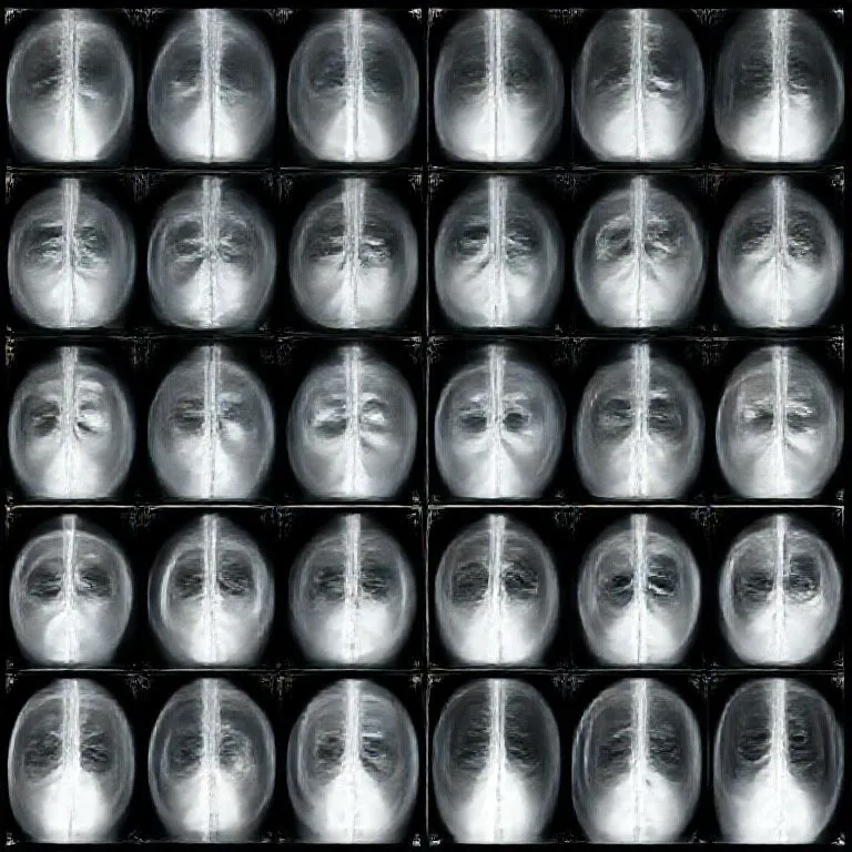
Brief description
An X-ray image is a shadow map of how much different materials absorb X-rays. Dense materials like bone or metal block more radiation and appear lighter, while soft tissue lets more through and appears darker.
Use / Function
- Medical diagnosis: Bones, lungs, teeth, and foreign objects.
- Non-destructive inspection: Cracks and voids in metal parts or welds.
- Security screening: Hidden items in luggage or containers.
- Alignment and quality control: Internal fit checks without disassembly.
- Scale: Clinical, industrial, and field use.
Operating principle
- X-ray source: A X-ray tube emits high-energy photons.
- Attenuation: Materials absorb X-rays based on density and thickness.
- Detection: A detector or film converts the remaining beam into an image.
- Contrast control: Collimation and shielding reduce scatter.
- Exposure control: Time and intensity balance detail and dose.
How to create it
- Prepare the source: Use a stable X-ray apparatus with shielding.
- Collimate the beam: Narrow it to the area of interest.
- Position the object: Place it between source and detector.
- Capture the image: Record with film or a digital detector.
- Review and adjust: Repeat with corrected exposure if needed.
Required technological level
Advanced. High voltage, vacuum components, and radiation safety procedures are required.
Materials needed
- Essential materials: Glass, Tungsten, Copper for conductors, Lead for shielding, Steel for structure.
- Core equipment: X-ray apparatus, X-ray tube, Vacuum Tube components.
- Thermal control: X-ray cooling system for high duty cycles.
Variants and improvements
- Film radiography: Simple and portable but slower to process.
- Digital detectors: Faster capture with lower dose and easier storage.
- Contrast-enhanced imaging: Uses Contrast agent to highlight vessels and soft tissue.
- Microfocus imaging: High detail for small parts.
- Rotating anode systems: Higher power and shorter exposure times.
Limits and risks
- Radiation exposure: Requires strict shielding, distance, and time limits.
- Soft tissue contrast: Limited without advanced techniques.
- Contrast reactions: Some agents can cause adverse reactions or kidney strain.
- Motion blur: Movement reduces sharpness.
- Scatter artifacts: Poor collimation degrades image quality.
Related materials
- X-ray: The imaging technique and basic setup.
- X-ray apparatus: Complete system for safe imaging.
- X-ray tube: The radiation source.
- Contrast agent: Improves visibility of soft tissue and vessels.
- Lead: Primary shielding material.
- Glass: Envelope material for vacuum components.