Generated with AI
X-ray apparatus
Brief description
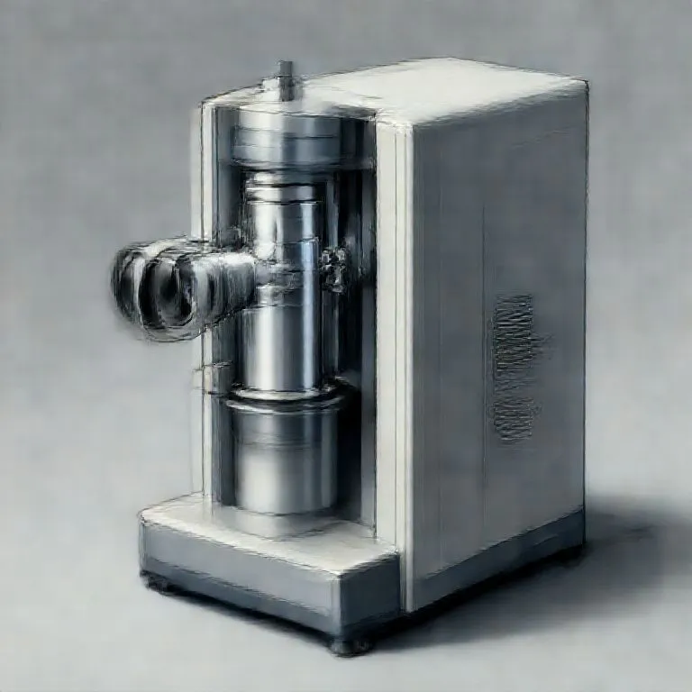
An X-ray apparatus is the complete system that generates, shapes, and records X-ray images. It combines the X-ray tube, shielding, power supply, and detector into a safe, usable imaging setup.
Use / Function
- Medical imaging: Radiography of bones, lungs, and dental structures.
- Inspection: Non-destructive testing of welds, castings, and assemblies.
- Security: Screening for hidden objects in luggage or containers.
- Alignment: Checking internal fit without disassembly.
Operating principle
- X-ray source: A Vacuum Tube emits X-rays when accelerated electrons strike a tungsten target.
- Beam shaping: Collimation narrows the beam to the area of interest.
- Object interaction: Dense materials absorb more X-rays, creating contrast.
- Detection: A detector or film captures the shadow image.
- Shielding: Lead-lined housing blocks stray radiation.
How to create it
- Assemble the tube: Seal a glass envelope with tungsten cathode and target.
- Provide power: Use a stable high-voltage supply for electron acceleration.
- Build the housing: Create a steel chassis with lead shielding.
- Add collimation: Include a narrow window or collimator for beam control.
- Install the detector: Mount film or a digital detector opposite the source.
Minimum functional version: a sealed tube, basic high-voltage supply, simple collimation, and film. Technical level is advanced due to vacuum work, high voltage, and radiation safety.
Materials needed
- Essential: Glass for the tube, Tungsten for filament/target, Copper for conductors.
- Shielding: Lead to block stray radiation.
- Structure: Steel for the chassis and mounts.
- Tools: Vacuum pump, high-voltage insulation, alignment tools.
Variants and improvements
- Rotating anode: Higher power with better heat distribution.
- Digital systems: Faster capture with lower dose and instant review.
- Portable units: Battery-powered field imaging.
- Fluoroscopy: Real-time imaging at higher technical complexity.
Limits and risks
- Radiation exposure: Requires strict shielding, distance, and time limits.
- High voltage: Severe electrical hazard during assembly and maintenance.
- Heat load: Targets overheat without cooling.
- Image limits: Scatter and motion blur reduce detail.